Baby mri scan tube information
Home » Trending » Baby mri scan tube informationYour Baby mri scan tube images are available. Baby mri scan tube are a topic that is being searched for and liked by netizens now. You can Get the Baby mri scan tube files here. Get all free photos and vectors.
If you’re looking for baby mri scan tube pictures information related to the baby mri scan tube interest, you have pay a visit to the ideal blog. Our website always provides you with suggestions for refferencing the highest quality video and picture content, please kindly search and locate more enlightening video content and images that fit your interests.
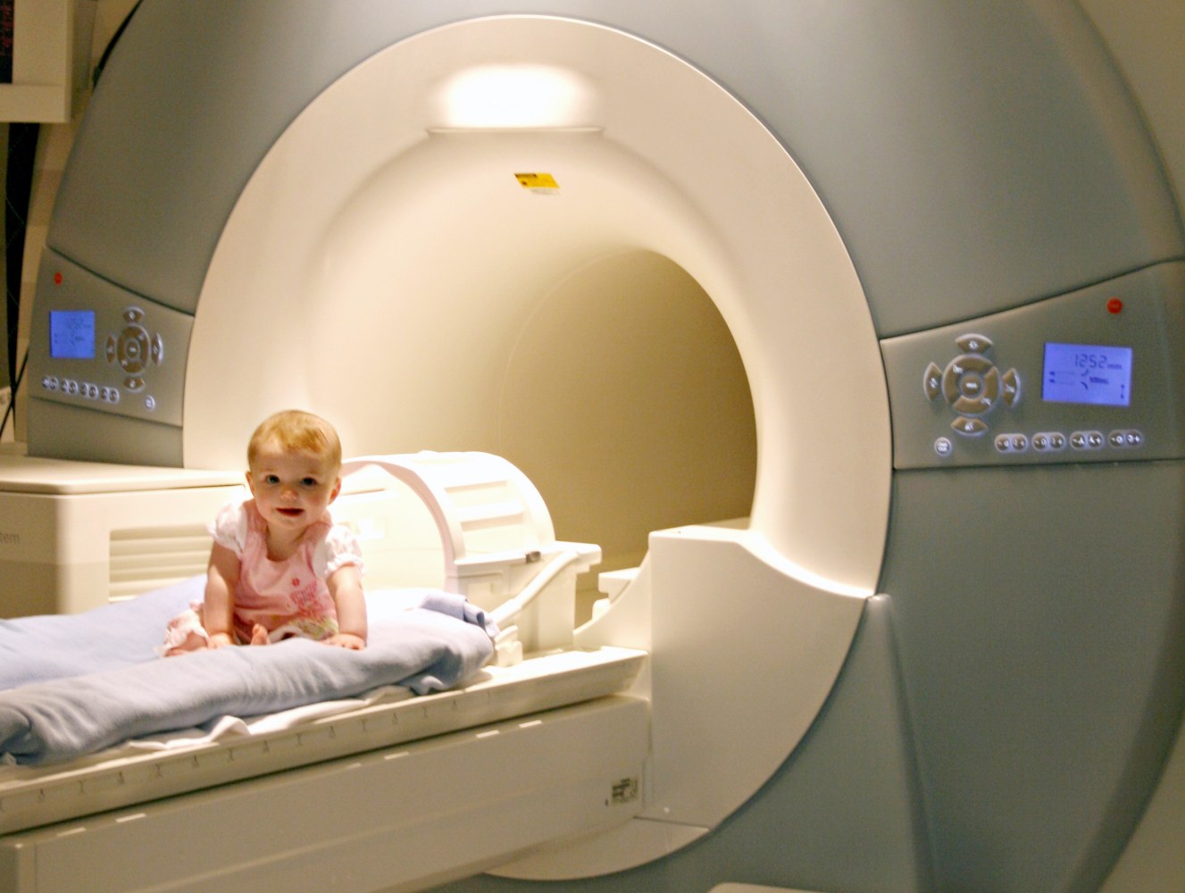
Baby Mri Scan Tube. The study published in the january 2017 issue of neurology, the journal from the american academy of neurology, found that performing an mri scan of a premature baby’s brain soon after birth can reveal areas of “white matter” in the brain, which is predictive of later disabilities. Once you’re in, the radiologist will leave the room and run the scan. Your baby will be prepared by the neonatal nursing team for their mri scan. A magnet surrounds the tube.
 Brain Scans Detect Signs of Autism in HighRisk Babies From
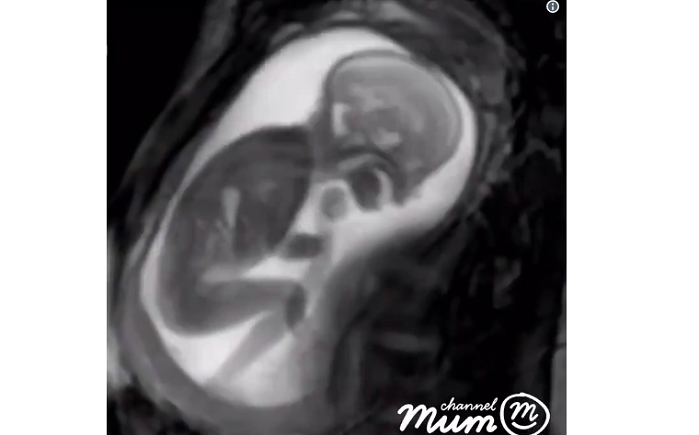
She seems to be trying to get comfortable. Once you’re in, the radiologist will leave the room and run the scan. Magnetic resonance imaging (mri) is a test done with a large machine that uses a magnetic field and pulses of radio wave energy to make pictures of organs and structures inside the belly. It uses strong magnetic fields and radio waves to produce a detailed image of organs and structures inside the body. Several expectant mothers had volunteered to participate in the experiment and five more births would be imaged with an mri machine. Breastfeeding your baby after an mri or ct scan this fact sheet aims to provide you with information about intravenous contrast mediums (imaging dyes) that may be used during your ct, mri or vq scan and help you make informed decisions about breastfeeding after your scan.
Your baby will be prepared by the neonatal nursing team for their mri scan.
Magnetic resonance imaging (mri) is a type of scan that uses strong magnetic fields and radio waves to produce detailed images of the inside of the body. A single mr image, like this one, takes several minutes to. Your baby will be prepared by the neonatal nursing team for their mri scan. Magnetic resonance imaging (mri) is a safe and painless test that uses a magnetic field and radio waves to produce detailed pictures of the body�s organs and structures. The analysis could also be helpful to plan the next pregnancy. A typical mri machine is a large tube with a hole at both ends.
 Source: vox.com
When brain damage is suspected in a newborn baby, brain scans are crucial for diagnosing and treating the injuries. If the baby can’t come to the mri, can the mri come to the baby? These images help in diagnosing a variety of conditions, from torn ligaments to tumours. Magnetic resonance imaging (mri) is a safe and painless test that uses a magnetic field and radio waves to produce detailed pictures of the body�s organs and structures. You may lie headfirst or feet first, depending on which part of your body your doctor wants to see.
 Source: smithsonianmag.com
You lie inside the tube during the scan. Ultrasound is used instead, which is much cheaper, more portable and convenient, but does not give as clear a picture as mri. An mri scan can be used to examine almost any part of the body, including the: A single mr image, like this one, takes several minutes to. How will my baby keep still during the mri scan?
 Source: en.mogaznews.com
A single mr image, like this one, takes several minutes to. These images help in diagnosing a variety of conditions, from torn ligaments to tumours. You may have to do it twice. Several expectant mothers had volunteered to participate in the experiment and five more births would be imaged with an mri machine. The analysis could also be helpful to plan the next pregnancy.
 Source: radiologykey.com
The study published in the january 2017 issue of neurology, the journal from the american academy of neurology, found that performing an mri scan of a premature baby’s brain soon after birth can reveal areas of “white matter” in the brain, which is predictive of later disabilities. The analysis could also be helpful to plan the next pregnancy. Brain scans for assessing risk of cerebral palsy, developmental delays. An mri differs from a cat scan (also called a ct scan or a computed axial tomography scan) because it doesn�t use radiation. The incredible connection between a mother and her baby is hard to put into words.
 Source:
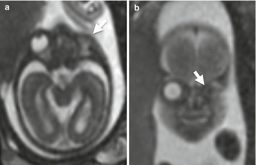
If other malformations are also present with anencephaly, doctors may order cytogenetic analysis to understand the exact cause. You will be asked to remain very still inside the machine during this process. The scanner will create strong magnetic. These images help in diagnosing a variety of conditions, from torn ligaments to tumours. A typical mri machine is a large tube with a hole at both ends.
 Source: doctorlib.info
Contrast mediums are usually inserted into your Breastfeeding your baby after an mri or ct scan this fact sheet aims to provide you with information about intravenous contrast mediums (imaging dyes) that may be used during your ct, mri or vq scan and help you make informed decisions about breastfeeding after your scan. An mri scan can be used to examine almost any part of the body, including the: A magnet surrounds the tube. During a magnetic resonance imaging scan, you’ll be asked to lie on a flat surface that’s slid into a scanner.
 Source: youtube.com
These images help in diagnosing a variety of conditions, from torn ligaments to tumours. You may lie headfirst or feet first, depending on which part of your body your doctor wants to see. Mri scans vary in the use of the medium to capture the imaging. She seems to be trying to get comfortable. You lie inside the tube during the scan.
 Source:
Contrast mediums are usually inserted into your Brain scans for assessing risk of cerebral palsy, developmental delays. A typical mri machine is a large tube with a hole at both ends. You may lie headfirst or feet first, depending on which part of your body your doctor wants to see. You lie inside the tube during the scan.
 Source:
She seems to be trying to get comfortable. They will need to be undressed (left in their nappy) and swaddled in a blanket, for warmth and security, for the scan. Magnetic resonance imaging (mri) and cranial ultrasonography (cus) are head imaging techniques, often called brain scans, that give doctors pictures of the baby’s brain. If the baby can’t come to the mri, can the mri come to the baby? A typical mri machine is a large tube with a hole at both ends.
 Source: verywellhealth.com
You lie inside the tube during the scan. Once you’re in, the radiologist will leave the room and run the scan. Magnetic resonance imaging (mri) is a type of scan that uses strong magnetic fields and radio waves to produce detailed images of the inside of the body. You may lie headfirst or feet first, depending on which part of your body your doctor wants to see. A typical mri machine is a large tube with a hole at both ends.
 Source:
Baby mri scan tube.and even though the 3t scanner has a larger bore, it doesn’t sacrifice quality like the open mri. An mri scan can be used to examine almost any part of the body, including the: Contrast mediums are usually inserted into your The analysis could also be helpful to plan the next pregnancy. Breastfeeding your baby after an mri or ct scan this fact sheet aims to provide you with information about intravenous contrast mediums (imaging dyes) that may be used during your ct, mri or vq scan and help you make informed decisions about breastfeeding after your scan.
 Source:
Ultrasound is used instead, which is much cheaper, more portable and convenient, but does not give as clear a picture as mri. The scanner will create strong magnetic. An mri uses strong magnetic fields and radio. If other malformations are also present with anencephaly, doctors may order cytogenetic analysis to understand the exact cause. These images help in diagnosing a variety of conditions, from torn ligaments to tumours.
 Source: huffingtonpost.co.uk
An mri uses strong magnetic fields and radio. Mri may be used to help diagnose or monitor treatment for a variety of conditions within the brain, chest, abdomen, pelvis and extremities. A baby drifts to sleep in the arms of his mother. It uses strong magnetic fields and radio waves to produce a detailed image of organs and structures inside the body. Brain damage in a baby:
 Source: lifenews.com
You lie inside the tube during the scan. How will my baby keep still during the mri scan? It uses strong magnetic fields and radio waves to produce a detailed image of organs and structures inside the body. Once you’re in, the radiologist will leave the room and run the scan. Mri scans vary in the use of the medium to capture the imaging.
 Source: internationalinside.com
Additionally, with a larger bore size and shorter tube, 3t mri patients don’t feel as enclosed. When brain damage is suspected in a newborn baby, brain scans are crucial for diagnosing and treating the injuries. You may lie headfirst or feet first, depending on which part of your body your doctor wants to see. Children�s magnetic resonance imaging (mri) uses a powerful magnetic field, radio waves and a computer to produce detailed pictures of the inside of your child�s body. Magnetic resonance imaging (mri) and cranial ultrasonography (cus) are head imaging techniques, often called brain scans, that give doctors pictures of the baby’s brain.
 Source: bio5.org
The intimate scenario is caught. Breastfeeding your baby after an mri or ct scan this fact sheet aims to provide you with information about intravenous contrast mediums (imaging dyes) that may be used during your ct, mri or vq scan and help you make informed decisions about breastfeeding after your scan. Once you’re in, the radiologist will leave the room and run the scan. Brain damage in a baby: She seems to be trying to get comfortable.
 Source:
You lie on a table that slides all the way into the tube. It uses strong magnetic fields and radio waves to produce a detailed image of organs and structures inside the body. You will be asked to remain very still inside the machine during this process. Mri may be used to help diagnose or monitor treatment for a variety of conditions within the brain, chest, abdomen, pelvis and extremities. A postnatal mri scan is often done to look for brain abnormalities.
 Source: nos.nl
When brain damage is suspected in a newborn baby, brain scans are crucial for diagnosing and treating the injuries. Ultrasound is used instead, which is much cheaper, more portable and convenient, but does not give as clear a picture as mri. Magnetic resonance imaging (mri) and cranial ultrasonography (cus) are head imaging techniques, often called brain scans, that give doctors pictures of the baby’s brain. The scanner will create strong magnetic. The study published in the january 2017 issue of neurology, the journal from the american academy of neurology, found that performing an mri scan of a premature baby’s brain soon after birth can reveal areas of “white matter” in the brain, which is predictive of later disabilities.
This site is an open community for users to do submittion their favorite wallpapers on the internet, all images or pictures in this website are for personal wallpaper use only, it is stricly prohibited to use this wallpaper for commercial purposes, if you are the author and find this image is shared without your permission, please kindly raise a DMCA report to Us.
If you find this site convienient, please support us by sharing this posts to your own social media accounts like Facebook, Instagram and so on or you can also bookmark this blog page with the title baby mri scan tube by using Ctrl + D for devices a laptop with a Windows operating system or Command + D for laptops with an Apple operating system. If you use a smartphone, you can also use the drawer menu of the browser you are using. Whether it’s a Windows, Mac, iOS or Android operating system, you will still be able to bookmark this website.
