Down syndrome baby at 14 weeks ultrasound Idea
Home » Trending » Down syndrome baby at 14 weeks ultrasound IdeaYour Down syndrome baby at 14 weeks ultrasound images are available in this site. Down syndrome baby at 14 weeks ultrasound are a topic that is being searched for and liked by netizens now. You can Get the Down syndrome baby at 14 weeks ultrasound files here. Download all royalty-free vectors.
If you’re looking for down syndrome baby at 14 weeks ultrasound images information linked to the down syndrome baby at 14 weeks ultrasound interest, you have come to the ideal blog. Our website always provides you with suggestions for seeking the highest quality video and image content, please kindly hunt and locate more enlightening video articles and graphics that fit your interests.
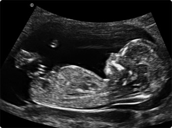
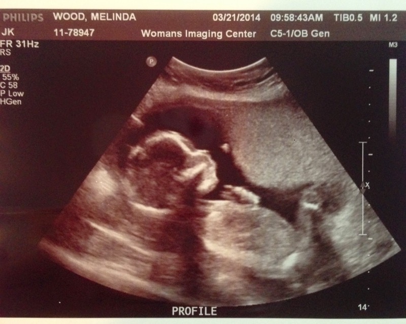
Down Syndrome Baby At 14 Weeks Ultrasound. Hi i am writing this post because baby centre was fantastic as a support network by reading everyones posts while i waited for my results from my percept test (harmony test) to return and i would like to share my happy story. A thicker neck can be an indicator of increased risk for birth defects such as down syndrome or trisomy 18. Between 11 and 14 weeks the nuchal translucency can be measured during an ultrasound examination. The results of the blood test, the nuchal translucency measurement and the mother�s age are used to estimate the risk for down syndrome and trisomy 18.
 4d Ultrasound Of Baby With Down Syndrome Captions Todays From captionstodaysnl.blogspot.com
This assessment is for women who wish to have a screening test for the prediction of a baby with down�s syndrome. Between 11 and 14 weeks the nuchal translucency can be measured during an ultrasound examination. A detailed anomaly scan done at 20 weeks can only detect 50% of down syndrome cases. This causes a wide range of both physical disability and learning difficulties. A thicker neck can be an indicator of increased risk for birth defects such as down syndrome or trisomy 18. Have had ultrasound and only one baby.
An ultrasound can detect fluid at the back of a fetus�s neck, which sometimes indicates down syndrome.
I am so excited that i get to post this. Between 11 and 14 weeks the nuchal translucency can be measured during an ultrasound examination. When the results of this blood test are combined with the results from the first trimester blood test and nuchal. The second step is a maternal blood test between 15 to 20 weeks of pregnancy. The developing baby is almost ready for birth and is now able to be seen in even more detail. However, ultrasound is often used as a screening test for down syndrome and other chromosome abnormalities.

You should take a screening test for risk assessment in order to prevent down syndrome pregnancy complications. To detect the chance of down syndrome, the 2 types of tests are combined based on the mother’s age. Checks for down syndrome, heart defects and other chromosomal abnormalities. Down syndrome ultrasound pictures 20 weeks. No nasal bone via ultrasound at 14 weeks:

The results of the blood test, the nuchal translucency measurement and the mother�s age are used to estimate the risk for down syndrome and trisomy 18. The results of the blood test, the nuchal translucency measurement and the mother�s age are used to estimate the risk for down syndrome and trisomy 18. A thicker neck can be an indicator of increased risk for birth defects such as down syndrome or trisomy 18. Because a baby�s nuchal translucency normally gets a bit thicker with each day of gestation, researchers have been able to establish how large the translucent area should be each day during the three weeks the. No nasal bone via ultrasound at 14 weeks:
 Source:
An ultrasound scan could save many mothers the decision over whether to have an amniocentesis and risk losing a baby. This is done to measure the thickness of fluid behind the baby’s neck, called nuchal translucency. The results of the blood test, the nuchal translucency measurement and the mother�s age are used to estimate the risk for down syndrome and trisomy 18. Because a baby�s nuchal translucency normally gets a bit thicker with each day of gestation, researchers have been able to establish how large the translucent area should be each day during the three weeks the. Family medicine 14 years experience.
 Source: co.pinterest.com
When the results of this blood test are combined with the results from the first trimester blood test and nuchal. A detailed anomaly scan done at 20 weeks can only detect 50% of down syndrome cases. Family medicine 14 years experience. The first was the nuchal translucency scan, which determines whether or not the baby appears to have down syndrome. Ultrasound of baby with down syndrome.

3d 4d ultrasound at 12 weeks pregnant. This assessment is for women who wish to have a screening test for the prediction of a baby with down�s syndrome. An ultrasound scan could save many mothers the decision over whether to have an amniocentesis and risk losing a baby. Have had ultrasound and only one baby. The developing baby is almost ready for birth and is now able to be seen in even more detail.

The test combines the woman�s age, the measurement of 2 proteins (biochemical markers) in the mother�s blood and a measurement of the skinfold behind the baby�s neck at 11 and a half to 14 weeks� of pregnancy. This measures the fluid under the skin at the back of the baby�s neck and can be used to determine your risk of having a baby with down syndrome. Down syndrome ultrasound pictures 20 weeks. The first was the nuchal translucency scan, which determines whether or not the baby appears to have down syndrome. Certain findings (sometimes called soft markers) on ultrasound may make your doctor more suspicious that your baby may have down syndrome.
 Source: themighty.com
An ultrasound scan could save many mothers the decision over whether to have an amniocentesis and risk losing a baby. However, ultrasound is often used as a screening test for down syndrome and other chromosome abnormalities. When the results of this blood test are combined with the results from the first trimester blood test and nuchal. Checks for down syndrome, heart defects and other chromosomal abnormalities. Mine were around 245, 000 at 9.2 weeks.
 Source:
When the results of this blood test are combined with the results from the first trimester blood test and nuchal. With the second trimester mss approximately 70% of down syndrome pregnancies can be detected. It�s called the combined test because it combines an ultrasound scan with a blood test. This week, we had two scheduled ultrasounds. If done between 10 and 13 weeks pregnant, the blood test and ultrasound scan together will detect around 90% of babies affected with down syndrome.
 Source: ldwtanka.blogspot.com
The ultrasound is called a nuchal translucency (nt) test and can be performed when you are between 11 to 14 weeks pregnant. Between 11 and 14 weeks the nuchal translucency can be measured during an ultrasound examination. However, ultrasound is often used as a screening test for down syndrome and other chromosome abnormalities. This assessment is for women who wish to have a screening test for the prediction of a baby with down�s syndrome. Hi i am writing this post because baby centre was fantastic as a support network by reading everyones posts while i waited for my results from my percept test (harmony test) to return and i would like to share my happy story.
 Source: youtube.com
Down syndrome ultrasound pictures 20 weeks. This is an effective way of down syndrome detection. This week, we had two scheduled ultrasounds. At the moment there still isn’t a completely safe test that will tell you that your baby definitely does or doesn’t have down’s syndrome, but the nhs offers everyone combined first trimester screening, which is a test performed at around 12 weeks using a combination of ultrasound scan findings and a basic. It�s called the combined test because it combines an ultrasound scan with a blood test.
 Source: nishiohmiya-golf.com
The ultrasound test is called measurement of nuchal translucency. For example, your likelihood of carrying a baby with down syndrome ranges from approximately 1 in 1,200 at age 25 to 1 in 100 at age 40. Have had ultrasound and only one baby. With the second trimester mss approximately 70% of down syndrome pregnancies can be detected. The developing baby is almost ready for birth and is now able to be seen in even more detail.
 Source:
You should take a screening test for risk assessment in order to prevent down syndrome pregnancy complications. It�s called the combined test because it combines an ultrasound scan with a blood test. A screening test for down�s syndrome, edwards� syndrome and patau�s syndrome is available between weeks 10 and 14 of pregnancy. For example, your likelihood of carrying a baby with down syndrome ranges from approximately 1 in 1,200 at age 25 to 1 in 100 at age 40. Could high levels of hcg point to down syndrome?
 Source: pinterest.com
This week, we had two scheduled ultrasounds. Down syndrome ultrasound pictures 20 weeks. This is an effective way of down syndrome detection. During the first trimester, this combined method results in more effective or comparable detection rates than methods used during the second trimester. The ultrasound examination cannot diagnose a fetus with down syndrome with certainty.
 Source:
However, ultrasound is often used as a screening test for down syndrome and other chromosome abnormalities. Only invasive tests (amniocentesis and chorionic villus sampling) can clinically confirm the presence of down syndrome in a baby. The developing baby is almost ready for birth and is now able to be seen in even more detail. If done between 10 and 13 weeks pregnant, the blood test and ultrasound scan together will detect around 90% of babies affected with down syndrome. An ultrasound scan could save many mothers the decision over whether to have an amniocentesis and risk losing a baby.
 Source: thebirthcompany.co.uk
The first was the nuchal translucency scan, which determines whether or not the baby appears to have down syndrome. Checks for down syndrome, heart defects and other chromosomal abnormalities. Could high levels of hcg point to down syndrome? This measures the fluid under the skin at the back of the baby�s neck and can be used to determine your risk of having a baby with down syndrome. O in fetuses with down syndrome, 6 (37%) of 16 did not have detectable nose bones compared with 1 (0.5%) of 223 control fetuses.
 Source:
Only invasive tests (amniocentesis and chorionic villus sampling) can clinically confirm the presence of down syndrome in a baby. The ultrasound examination cannot diagnose a fetus with down syndrome with certainty. Checks for down syndrome, heart defects and other chromosomal abnormalities. This is an effective way of down syndrome detection. This measures the fluid under the skin at the back of the baby�s neck and can be used to determine your risk of having a baby with down syndrome.
 Source: pinterest.com
For example, your likelihood of carrying a baby with down syndrome ranges from approximately 1 in 1,200 at age 25 to 1 in 100 at age 40. Also asked, can down syndrome be detected at 20 week ultrasound? A thicker neck can be an indicator of increased risk for birth defects such as down syndrome or trisomy 18. The ultrasound test is called measurement of nuchal translucency. This measures the fluid under the skin at the back of the baby�s neck and can be used to determine your risk of having a baby with down syndrome.
 Source: captionstodaysnl.blogspot.com
This is an effective way of down syndrome detection. When the results of this blood test are combined with the results from the first trimester blood test and nuchal. This assessment is for women who wish to have a screening test for the prediction of a baby with down�s syndrome. A thicker neck can be an indicator of increased risk for birth defects such as down syndrome or trisomy 18. This causes a wide range of both physical disability and learning difficulties.
This site is an open community for users to do submittion their favorite wallpapers on the internet, all images or pictures in this website are for personal wallpaper use only, it is stricly prohibited to use this wallpaper for commercial purposes, if you are the author and find this image is shared without your permission, please kindly raise a DMCA report to Us.
If you find this site convienient, please support us by sharing this posts to your own social media accounts like Facebook, Instagram and so on or you can also bookmark this blog page with the title down syndrome baby at 14 weeks ultrasound by using Ctrl + D for devices a laptop with a Windows operating system or Command + D for laptops with an Apple operating system. If you use a smartphone, you can also use the drawer menu of the browser you are using. Whether it’s a Windows, Mac, iOS or Android operating system, you will still be able to bookmark this website.
