How to see teeth xray Idea
Home » Trend » How to see teeth xray IdeaYour How to see teeth xray images are ready in this website. How to see teeth xray are a topic that is being searched for and liked by netizens today. You can Get the How to see teeth xray files here. Download all royalty-free vectors.
If you’re searching for how to see teeth xray pictures information related to the how to see teeth xray topic, you have pay a visit to the ideal blog. Our site frequently gives you suggestions for refferencing the maximum quality video and picture content, please kindly surf and locate more enlightening video articles and graphics that fit your interests.
How To See Teeth Xray. In between the teeth, the gums protect layers of bone. It can also detect anatomical abnormalities with the floor of the mouth or the palate. As an example, a study by uraba found that when evaluating teeth that due to anatomical considerations are characteristically more difficult to examine radiographically (upper incisors, canines and molars), that the use of cbct was able to identify signs of an endodontic problem (periapical lesions, see pictures above) in 20% more cases. This includes both the lower and upper jaws, as well as all surrounding tissues and structures.
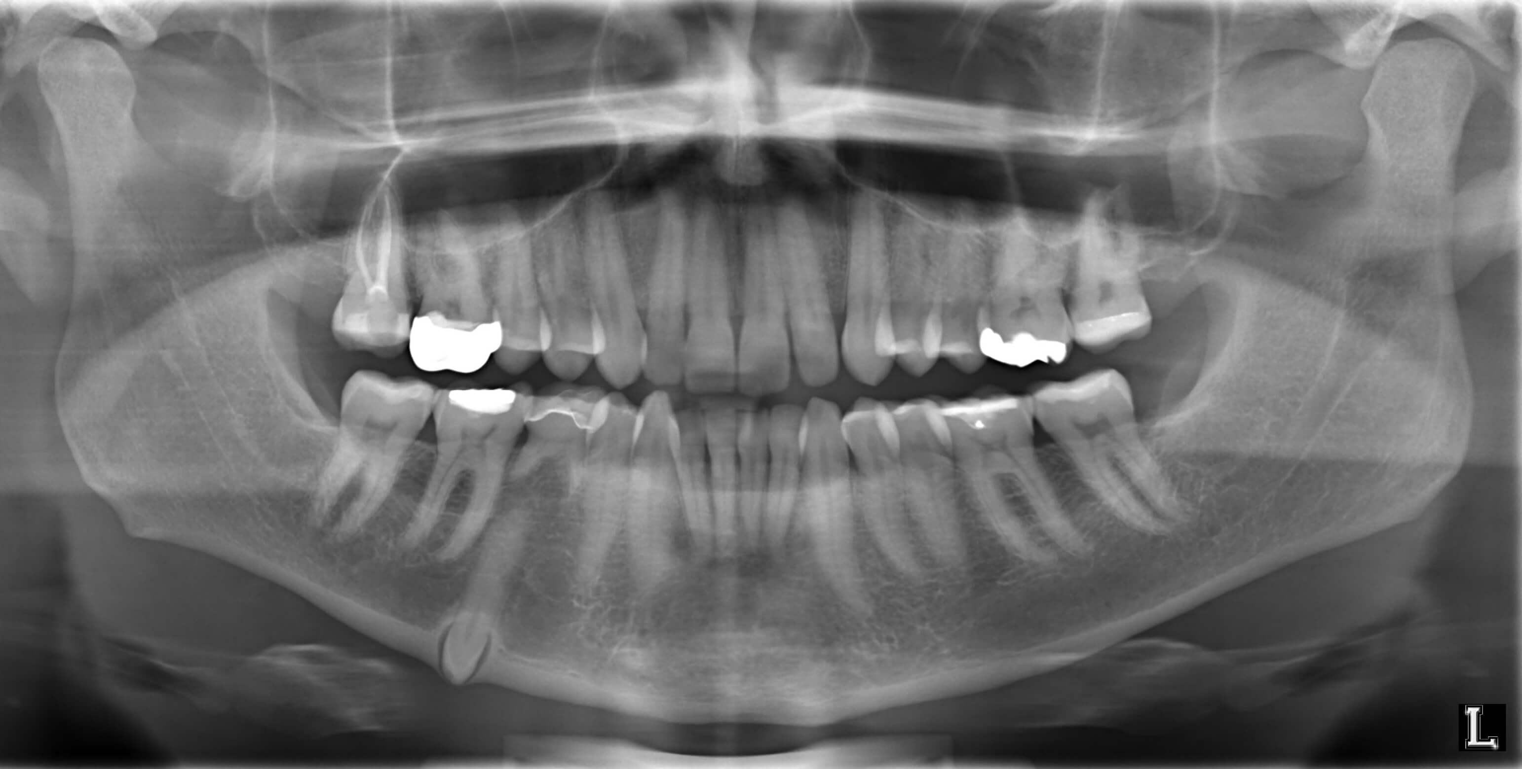 How Often Should Your Child Go For Regular Dental XRay From
Tomograms show a particular layer or “slice” of the mouth and blur out other layers. They allow the dentist to focus in on one specific tooth. That is a typical sign of an infection originating from the tooth�s pulp. It can also detect anatomical abnormalities with the floor of the mouth or the palate. The panorex enables surgeons to diagnose accurately, plan and safely treat wisdom teeth extractions. In most cases, it is used to visualise hard tissues like bones and joints but sometimes it is employed to capture soft tissues too.
That is a typical sign of an infection originating from the tooth�s pulp.
It is used to study the teeth in relation to the jaw and the person’s face profile. Wisdom teeth can be impacted by these photos, and a tumor can even be diagnosed using these photos. They allow the dentist to focus in on one specific tooth. The panorex shows the teeth and roots and their stages of development. In addition to showing all your teeth, the images also show your sinuses, jaw joints, and jaw bones as well. This includes both the lower and upper jaws, as well as all surrounding tissues and structures.
 Source:
No need to register, buy now! They allow the dentist to focus in on one specific tooth. Lateral cephalometric captures the entire side of the head. This includes both the lower and upper jaws, as well as all surrounding tissues and structures. In between the teeth, the gums protect layers of bone.
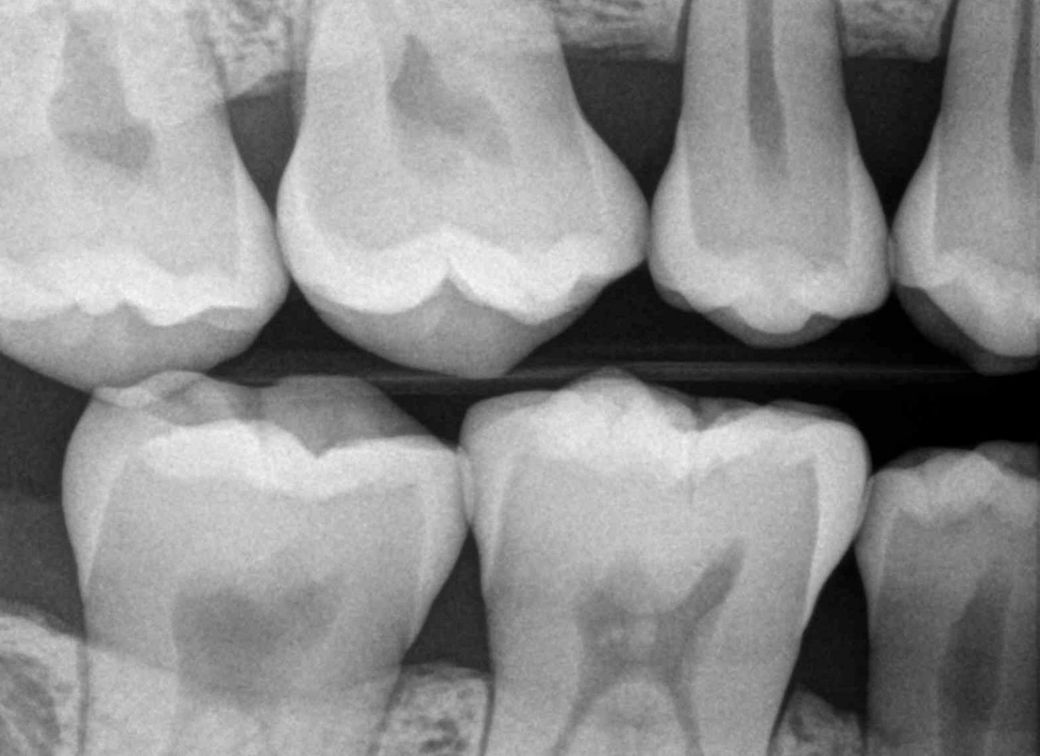 Source: seattlesmilesdental.com
Huge collection, amazing choice, 100+ million high quality, affordable rf and rm images. The panorex shows the teeth and roots and their stages of development. No need to register, buy now! It also shows their relationship to adjacent teeth, sinuses, and nerve canals. The panorex enables surgeons to diagnose accurately, plan and safely treat wisdom teeth extractions.
Source: quora.com
Find the perfect teeth xray stock photo. Wisdom teeth can be impacted by these photos, and a tumor can even be diagnosed using these photos. It can also detect anatomical abnormalities with the floor of the mouth or the palate. If you notice the second tooth from the left, you can see a dark aura surrounding the tip of the root. That is a typical sign of an infection originating from the tooth�s pulp.
 Source:
Point out how this portion protects the inner tooth. Find the perfect teeth xray stock photo. As an example, a study by uraba found that when evaluating teeth that due to anatomical considerations are characteristically more difficult to examine radiographically (upper incisors, canines and molars), that the use of cbct was able to identify signs of an endodontic problem (periapical lesions, see pictures above) in 20% more cases. It can also detect anatomical abnormalities with the floor of the mouth or the palate. In most cases, it is used to visualise hard tissues like bones and joints but sometimes it is employed to capture soft tissues too.
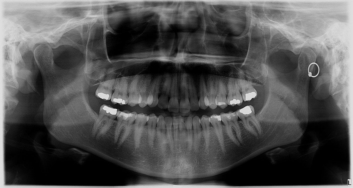 Source: sciencefriday.com
It can also detect anatomical abnormalities with the floor of the mouth or the palate. Some problems with the teeth are located deep inside the gums and structures and practically invisible to the naked eye. “while the cells that are necessary to form wisdom teeth are all present in the jaws at birth, they don�t start the process of forming the wisdom teeth until about the age of 7,” busaidy says. Wisdom teeth can be impacted by these photos, and a tumor can even be diagnosed using these photos. No need to register, buy now!
 Source: dentimax.com
Wisdom teeth can be impacted by these photos, and a tumor can even be diagnosed using these photos. This includes both the lower and upper jaws, as well as all surrounding tissues and structures. This is often the case when assessing the overall oral health of new patients, or when planning dental implant treatment. It also shows their relationship to adjacent teeth, sinuses, and nerve canals. As an example, a study by uraba found that when evaluating teeth that due to anatomical considerations are characteristically more difficult to examine radiographically (upper incisors, canines and molars), that the use of cbct was able to identify signs of an endodontic problem (periapical lesions, see pictures above) in 20% more cases.
 Source: dryazdan.com
Huge collection, amazing choice, 100+ million high quality, affordable rf and rm images. Some problems with the teeth are located deep inside the gums and structures and practically invisible to the naked eye. In most cases, it is used to visualise hard tissues like bones and joints but sometimes it is employed to capture soft tissues too. The panorex shows the teeth and roots and their stages of development. Wisdom teeth can be impacted by these photos, and a tumor can even be diagnosed using these photos.
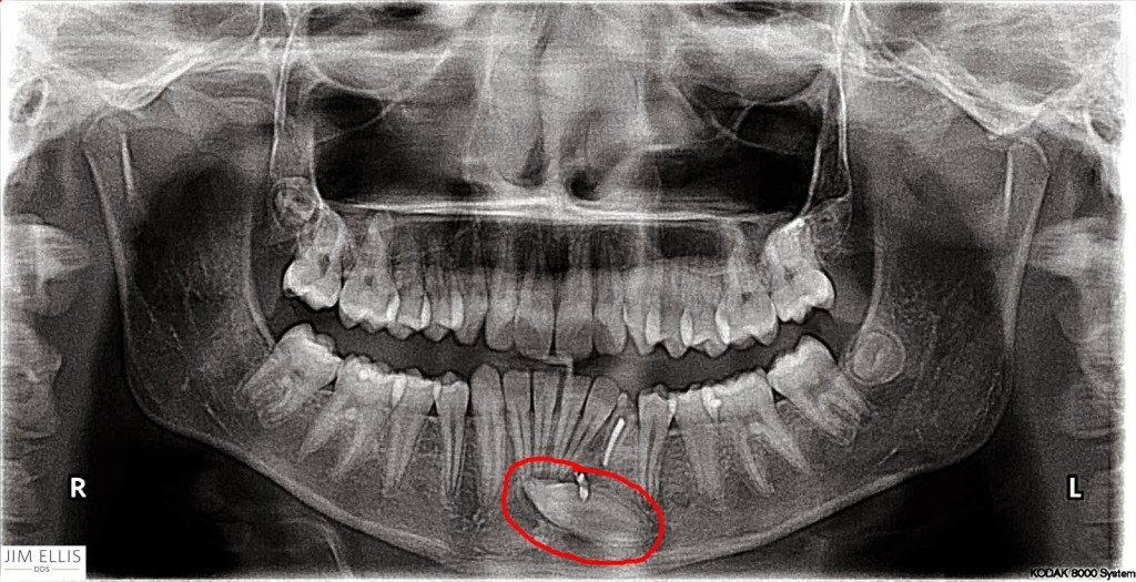 Source: bestogdendentist.com
That is a typical sign of an infection originating from the tooth�s pulp. This is often the case when assessing the overall oral health of new patients, or when planning dental implant treatment. Point out how this portion protects the inner tooth. In addition to showing all your teeth, the images also show your sinuses, jaw joints, and jaw bones as well. It can also detect anatomical abnormalities with the floor of the mouth or the palate.
 Source: pinterest.com
It can also detect anatomical abnormalities with the floor of the mouth or the palate. No need to register, buy now! They may also be used to see how the teeth touch each other when the patient bites down. If you notice the second tooth from the left, you can see a dark aura surrounding the tip of the root. Huge collection, amazing choice, 100+ million high quality, affordable rf and rm images.
 Source:
Huge collection, amazing choice, 100+ million high quality, affordable rf and rm images. That is a typical sign of an infection originating from the tooth�s pulp. Huge collection, amazing choice, 100+ million high quality, affordable rf and rm images. They may also be used to see how the teeth touch each other when the patient bites down. No need to register, buy now!
 Source: sciencephoto.com
It is used to study the teeth in relation to the jaw and the person’s face profile. Tomograms show a particular layer or “slice” of the mouth and blur out other layers. Some problems with the teeth are located deep inside the gums and structures and practically invisible to the naked eye. Point out how this portion protects the inner tooth. Wisdom teeth can be impacted by these photos, and a tumor can even be diagnosed using these photos.
 Source: canadianacademyofdentalhygiene.ca
They allow the dentist to focus in on one specific tooth. They may also be used to see how the teeth touch each other when the patient bites down. In most cases, it is used to visualise hard tissues like bones and joints but sometimes it is employed to capture soft tissues too. It is used to study the teeth in relation to the jaw and the person’s face profile. This alveolar bone contains sockets that hold teeth into place.
 Source: flickr.com
“while the cells that are necessary to form wisdom teeth are all present in the jaws at birth, they don�t start the process of forming the wisdom teeth until about the age of 7,” busaidy says. Huge collection, amazing choice, 100+ million high quality, affordable rf and rm images. This includes both the lower and upper jaws, as well as all surrounding tissues and structures. Point out how this portion protects the inner tooth. Wisdom teeth can be impacted by these photos, and a tumor can even be diagnosed using these photos.
 Source: greenspandental.com
They allow the dentist to focus in on one specific tooth. With a panorex single fmx, your dentist can view: Some problems with the teeth are located deep inside the gums and structures and practically invisible to the naked eye. As an example, a study by uraba found that when evaluating teeth that due to anatomical considerations are characteristically more difficult to examine radiographically (upper incisors, canines and molars), that the use of cbct was able to identify signs of an endodontic problem (periapical lesions, see pictures above) in 20% more cases. This alveolar bone contains sockets that hold teeth into place.
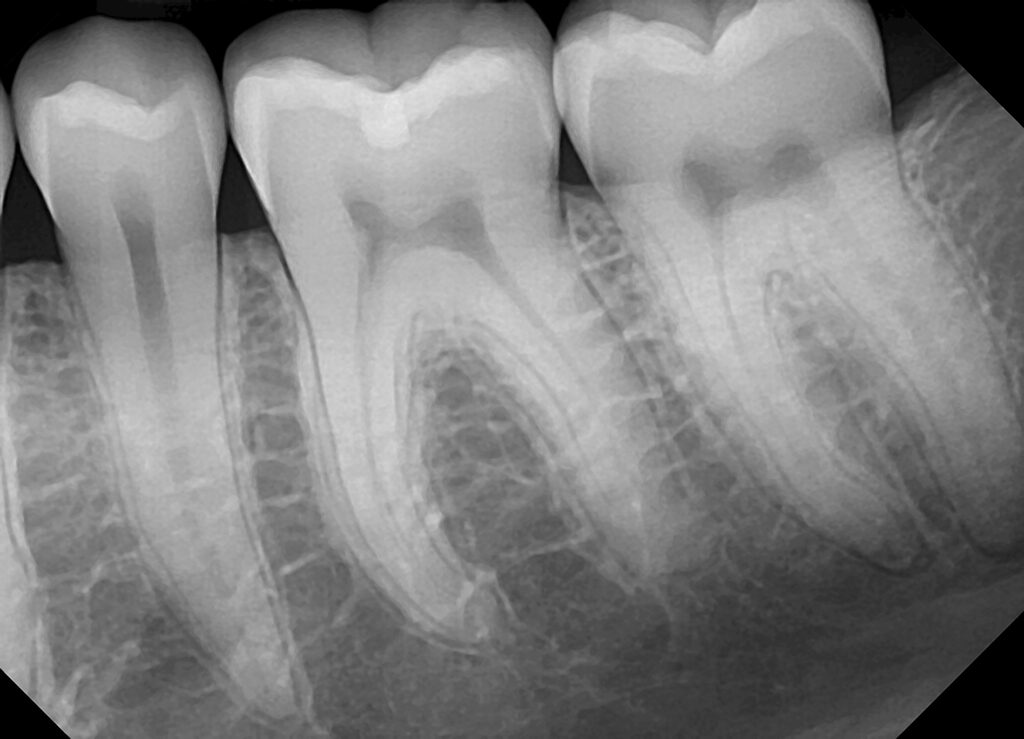 Source: dentimax.com
Lateral cephalometric captures the entire side of the head. They allow the dentist to focus in on one specific tooth. Wisdom teeth can be impacted by these photos, and a tumor can even be diagnosed using these photos. As an example, a study by uraba found that when evaluating teeth that due to anatomical considerations are characteristically more difficult to examine radiographically (upper incisors, canines and molars), that the use of cbct was able to identify signs of an endodontic problem (periapical lesions, see pictures above) in 20% more cases. It is used to study the teeth in relation to the jaw and the person’s face profile.
 Source: delacombefamilydental.com.au
It is used to study the teeth in relation to the jaw and the person’s face profile. This is often the case when assessing the overall oral health of new patients, or when planning dental implant treatment. If you notice the second tooth from the left, you can see a dark aura surrounding the tip of the root. In most cases, it is used to visualise hard tissues like bones and joints but sometimes it is employed to capture soft tissues too. In addition to showing all your teeth, the images also show your sinuses, jaw joints, and jaw bones as well.
 Source:
It can also detect anatomical abnormalities with the floor of the mouth or the palate. It is used to study the teeth in relation to the jaw and the person’s face profile. That is a typical sign of an infection originating from the tooth�s pulp. Find the perfect teeth xray stock photo. It can also detect anatomical abnormalities with the floor of the mouth or the palate.
 Source:
It can also detect anatomical abnormalities with the floor of the mouth or the palate. With a panorex single fmx, your dentist can view: If you notice the second tooth from the left, you can see a dark aura surrounding the tip of the root. In between the teeth, the gums protect layers of bone. As an example, a study by uraba found that when evaluating teeth that due to anatomical considerations are characteristically more difficult to examine radiographically (upper incisors, canines and molars), that the use of cbct was able to identify signs of an endodontic problem (periapical lesions, see pictures above) in 20% more cases.
This site is an open community for users to submit their favorite wallpapers on the internet, all images or pictures in this website are for personal wallpaper use only, it is stricly prohibited to use this wallpaper for commercial purposes, if you are the author and find this image is shared without your permission, please kindly raise a DMCA report to Us.
If you find this site serviceableness, please support us by sharing this posts to your preference social media accounts like Facebook, Instagram and so on or you can also bookmark this blog page with the title how to see teeth xray by using Ctrl + D for devices a laptop with a Windows operating system or Command + D for laptops with an Apple operating system. If you use a smartphone, you can also use the drawer menu of the browser you are using. Whether it’s a Windows, Mac, iOS or Android operating system, you will still be able to bookmark this website.
